


From Gene to Structure: Demonstrating Cryo-EM-Ready Protein Quality Using STEAP2 as a Model Target
Abstract
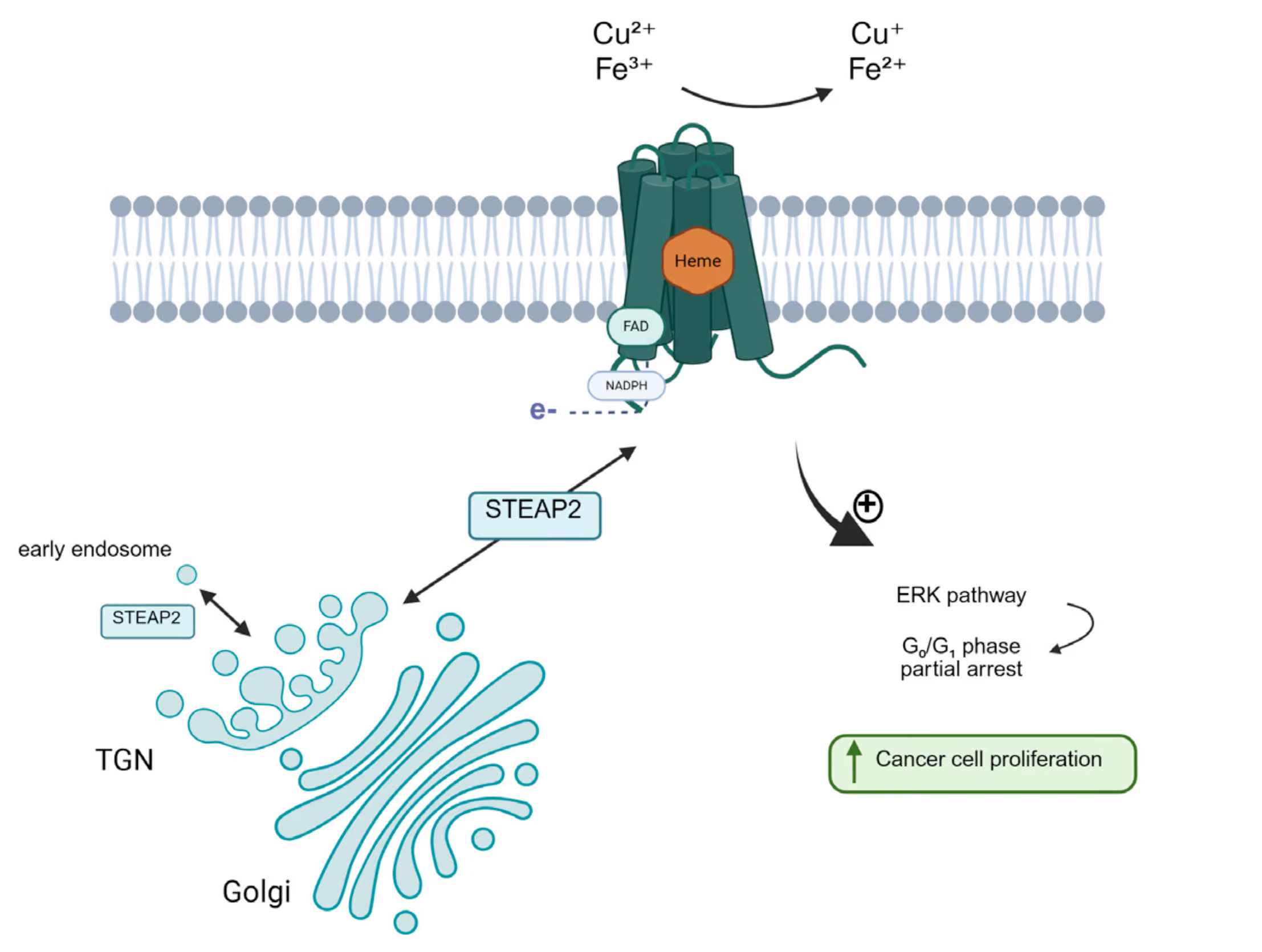
Six Transmembrane Epithelial Antigen of Prostate 2 (STEAP2) is a metalloreductase involved in transmembrane electron transfer, contributing to the uptake and regulation of iron and copper
ions. While its precise physiological role is still being investigated, STEAP2 is primarily expressed in prostate tissue and localized to the Golgi apparatus, tubular structures, and the plasma membrane (1–5) (Fig. 1). STEAP2 is overexpressed in invasive prostate cancer, where it promotes cell proliferation, migration, and invasion (2,6–8). In contrast, it is downregulated in breast cancer, where it may function as a tumor suppressor by inhibiting epithelial–mesenchymal transition (EMT) and PI3K/AKT signaling
(9). Abnormal copper levels are observed in various cancers,which may link STEAP2’s role in metal regulation of malignancy.In hepatocellular carcinoma, STEAP2 knockdown altered copperhomeostasis and reduced cancer cell migration and invasion
(1). These findings highlight STEAP2 as a potential therapeutic target across several cancer types, though its complete molecular function remains to be elucidated (10).
In this application note, STEAP2 is used to demonstrate Eurofins CALIXAR’s capability to produce native, high-quality membraneproteins suitable for high-resolution structural studies. Cryo-electron microscopy (cryo-EM) demands protein samples that are structurally homogeneous, stable, and monodisperse - criteria met only through a robust, carefully designed and optimized production and purification workflow. The successful structural determination of STEAP2 demonstrates the reliability of Eurofins CALIXAR’s process and its relevance to drug discovery efforts targeting complex membrane proteins.
To achieve this, Eurofins CALIXAR produced native STEAP2, now available as catalog product (#41-o101a) in human HEK293 cells using its proprietary expression and purification platform, ensuring the isolation of structurally intact, functionally relevant protein. In collaboration with Eyen SE, high-resolution cryo-M analysis of STEAP2 bound to NADP⁺, FAD⁺, and heme was performed, yielding a 3D structure at 2.1 Å resolution, showing a significant improvement over the previous 3.2 Å structure (PDB:7TAI) (11). The combination of high map quality, rapid timelines, and cost-effective workflow makes this approach ideally suited for late Hit-to-Lead and Lead Optimization stages, providing actionable structural insights to guide medicinal and synthetic chemistry programs.
Methods
Protein Construction & Expression
STEAP2 was cloned into a pOET6 vector featuring a C-terminal GFP-tag and a FLAG-TwinStrep tag for purification. An HRV-3C cleavage site was introduced N-terminal to the GFP for tag removal. Protein expression was carried out in Freestyle™ 293F cells via the BacMam system for 48 h at 37 °C in the presence of 10 mM sodium butyrate. Cells were harvested, lysed mechanically, and membranes were isolated by ultracentrifugation.
Protein Solubilization & SEC Purification
Membranes were solubilized at 5 mg/mL total protein using 20 mM DDM and 2 mM Xtab-DCOD (Eurofins CALIXAR). STEAP2 was purified using Strep-Tactin® affinity chromatography, followed by HRV-3C protease (Eurofins Calixar) cleavage to remove the tags. HRV-3C from Eurofins Calixar is a recombinant protease with unique strep tag permitting simple target protein re-purification processes. Final polishing was performed via size-exclusion chro-matography (SEC) on a Superose 6 Increase 10/300 GL column to obtain cleaved STEAP2 for cryo-EM.
Cryostability Assay
Cleaved STEAP2 underwent several freeze–thaw cycles. After each cycle, samples were analyzed on a NanoTemper Tycho instrument using the manufacturer’s protocols.
Heme Binding Assay
Spectroscopic analysis (340–600 nm) was used to confirm heme binding. A hemin-containing buffer was used as a negative control.
Cryo-EM Structure Determination
Initial sample quality was assessed on Talos Arctica and Glacios 2 electron microscopes, both equipped with Falcon 4i direct detectors. Around 200 micrographs were acquired on standard Quantifoil Cu 2/1 grids. STEAP2 particles were homogeneously distributed, with clear top and side views and well-defined transmembrane secondary structure in 2D class averages—ndicative of structural integrity, thin detergent micelles, and favorable vitrification properties. Other grid and support types were tested but did not outperform the default conditions. 2 High-resolution feasibility and final data collection were performed on a Titan Krios (300 kV) equipped with a Gatan K3 camera. Eyen’s internal processing pipeline, based on custom Relion and Scipion workflows, was used. An initial 3.5 Å map was obtained from ~700 movies, with clear density for NADP +, FAD+, and heme, and a favorable particle count-to-resolution curve. The final 2.1 Å structure was reconstructed from ~380,000 particles collected over 4600 movies in a 6-hour session. Starting from the 7TAI PDB structure, modeling included manual rebuilding based on the actual sequence and refinement with guided mo-lecular dynamics. Non-bonded interactions were considered to improve model quality. Binding interactions between STEAP2 residues and its ligands were further characterized.
STEAP2 Expression
A STEAP2 construct was engineered with C-terminal affinity tags (TwinStrep and FLAG) and a GFP moiety for convenient detection and purification. Transient expression in FreeStyle 293F cells was performed using the BacMAM system. To optimize yield and stability, expression was tested at multiple time points and temperatures. Maximum expression was achieved at 37°C after 48 hours post-transfection, while longer incubation led to degradation of the target protein (Figure 2). These results demonstrate that while the expression system is fundamentally reliable, producing high-quality yields of sensitive membrane proteins like STEAP2 requires careful optimization of conditions such as temperature and incubation time. Without this fine-tuning, prolonged expression can lead to protein degradation, compromising sample integrity and complicating downstream analyses. Effectively managing these variables is essential not only for maximizing protein production, but also for preserving the native structure and function of these fragile proteins—ultimately enabling more accurate biochemical and structural studies.
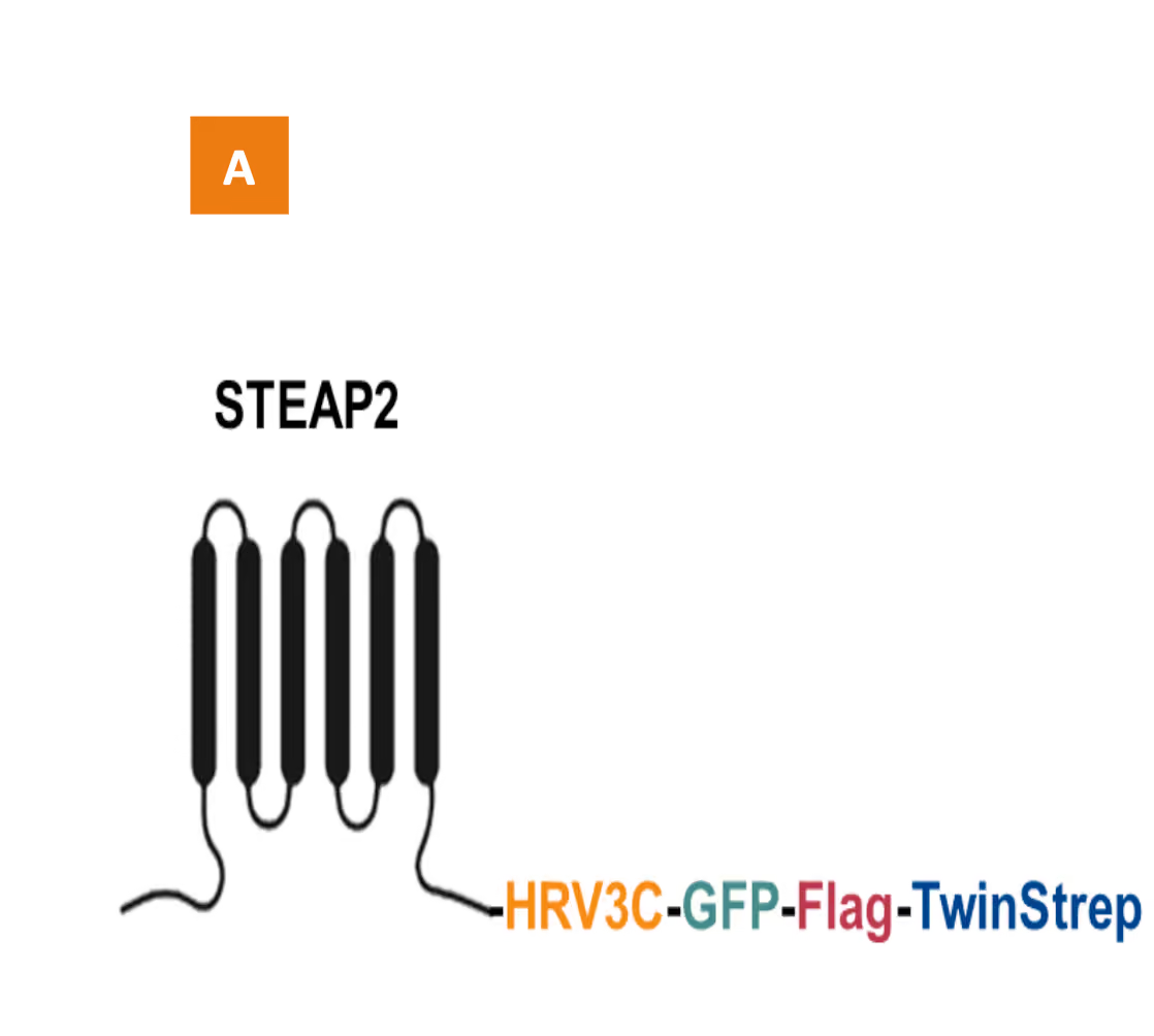
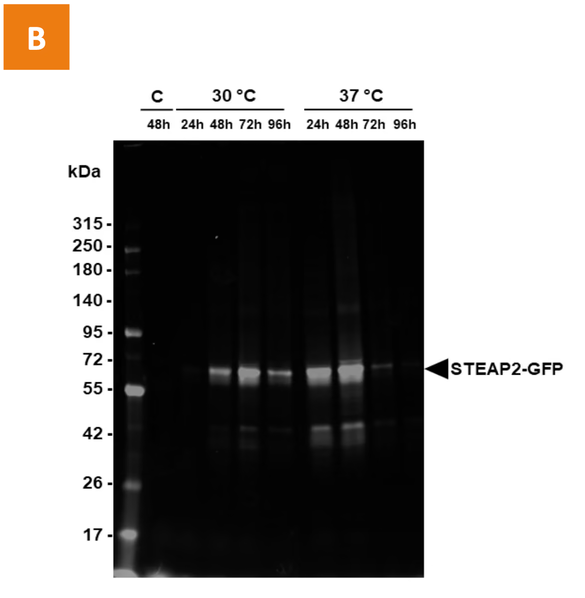
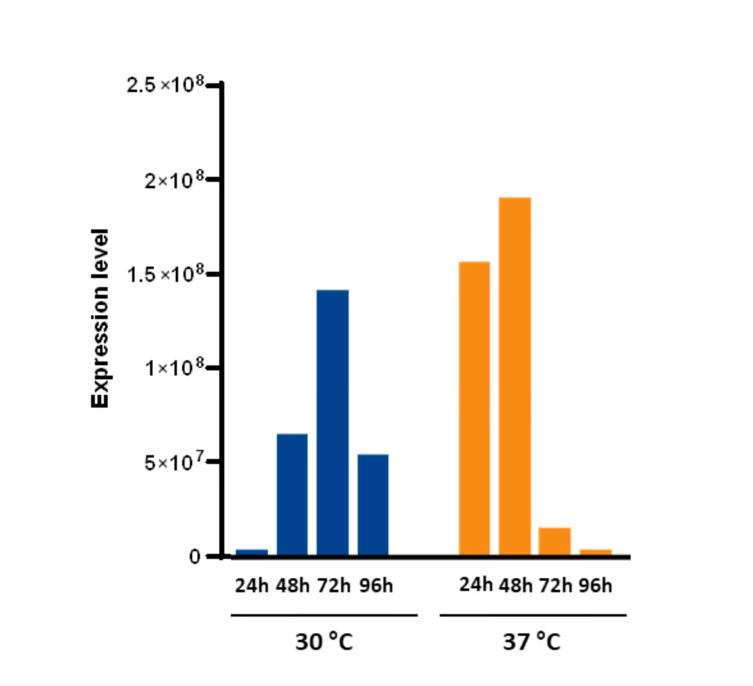
Figure 2. STEAP2 Expression. (A) Schematic representation of the STEAP2 construct with HRV-3C cleavage site, GFP, and dual affinity tags. (B) Expression profile
of STEAP2 in FreeStyle 293F cells. SDS-PAGE 4-15% GFP detection. Cells were harvested at 24, 48, 72, and 96 hours post-transfection. Protein levels were analyzed by
SDS-PAGE followed by in-gel GFP fluorescence detection. Quantified STEAP2-GFP signals are presented as a bar graph.
STEAP2 Solubilization & Purification
As part of its standard workflow, Eurofins CALIXAR evaluates membrane protein solubility and stability across a panel of detergents to identify optimal conditions for native extraction and purification. For STEAP2, membranes were solubilized using 20 mM DDM and 2 mM Xstab-DCOD, a Eurofins CALIXAR proprietary detergent known to stabilize membrane proteins. Purification of STEAP2 was performed using the C-terminal TwinStrep tag and a Strep-Tactin®XT 4Flow® resin on an FPLC system. Starting from 2 L of transfected FreeStyle 293F cells, the solubilized membrane protein was loaded onto a 4 mL column and eluted with a biotin-containing buffer. The resulting fractions contained pure STEAP2 (purity >80%), as demonstrated by total protein staining (Figure 3A) and GFP-specific in-gel fluorescence (Figure 3B).
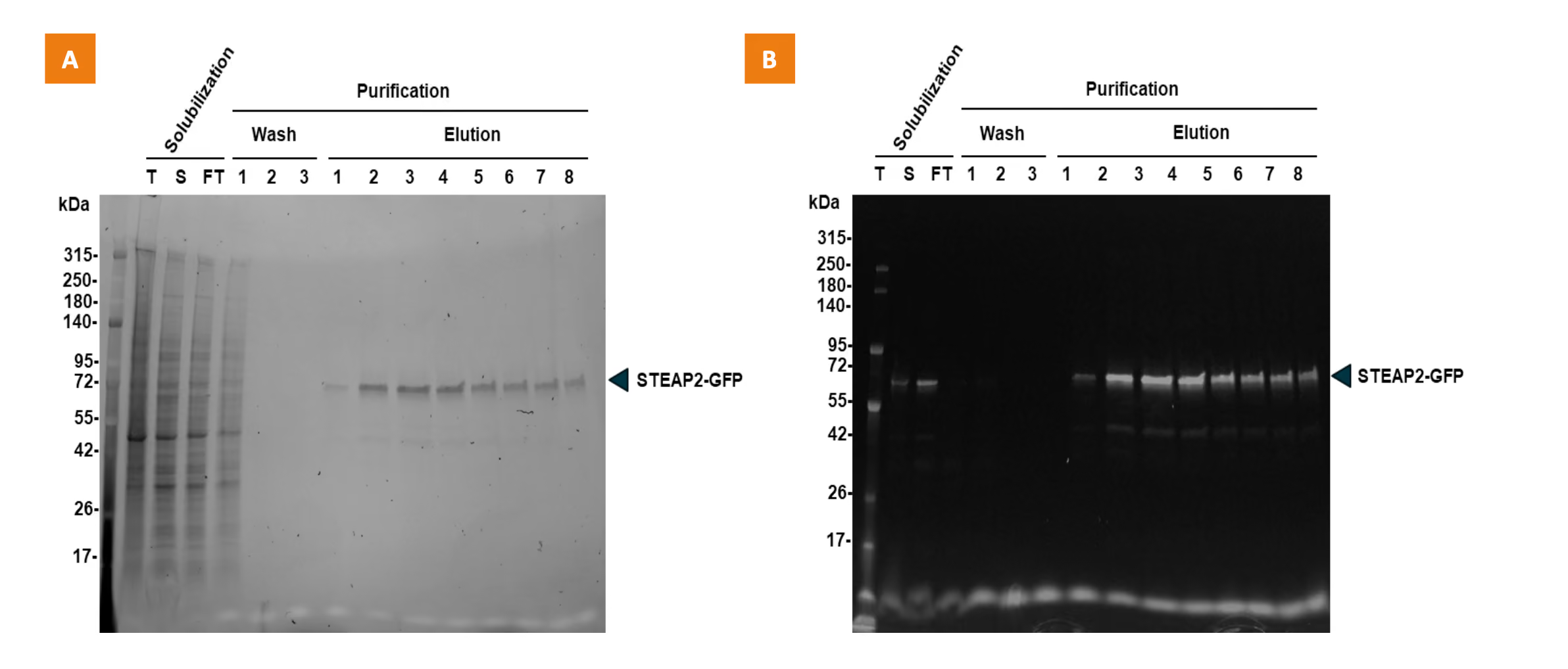
Figure 3. Affinity purification of STEAP2. (A) SDS-PAGE analysis of eluted fractions using Stain-Free™ total protein detection. (B) In-gel GFP fluorescence confirms specific detection of STEAP2-GFP.
Purified, Stable & Functional STEAP2
Affinity-purified STEAP2 appeared relatively pure by SDS-PAGE (Figure 4A). To evaluate sample preparation, chrono- and cryo-stability assays were performed to determine whether the sample could be maintained without degradation over time. No degradation or precipitation was detected after 72 hours at 4°C (Figure 4B), and the protein remained stable through three freeze-thaw cycles without a cryoprotectant, as shown by Native-PAGE (Figure 4C). This allowed the research team to verify the protein’s integrity and advance to more extensive biochemical and structural studies.
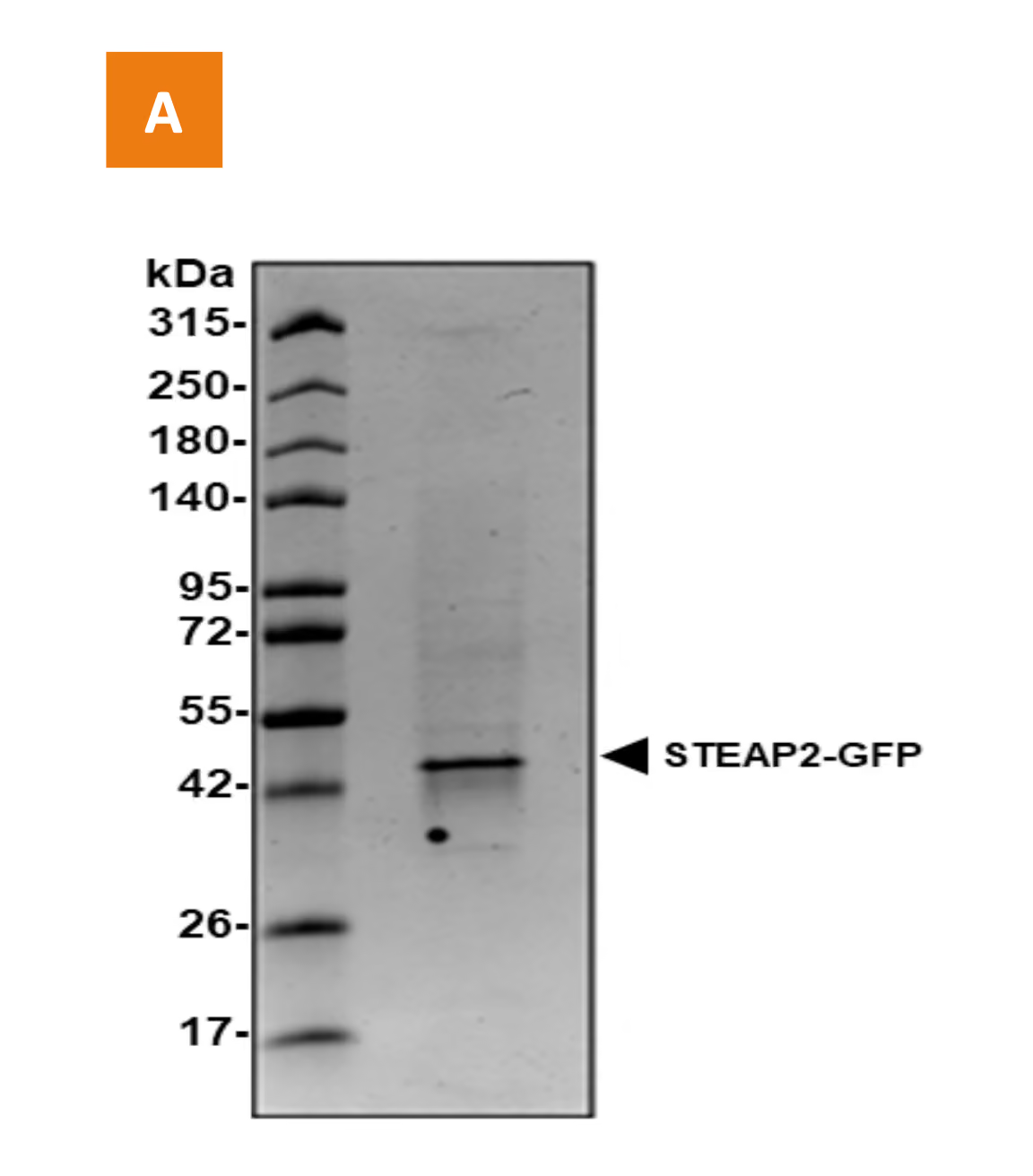
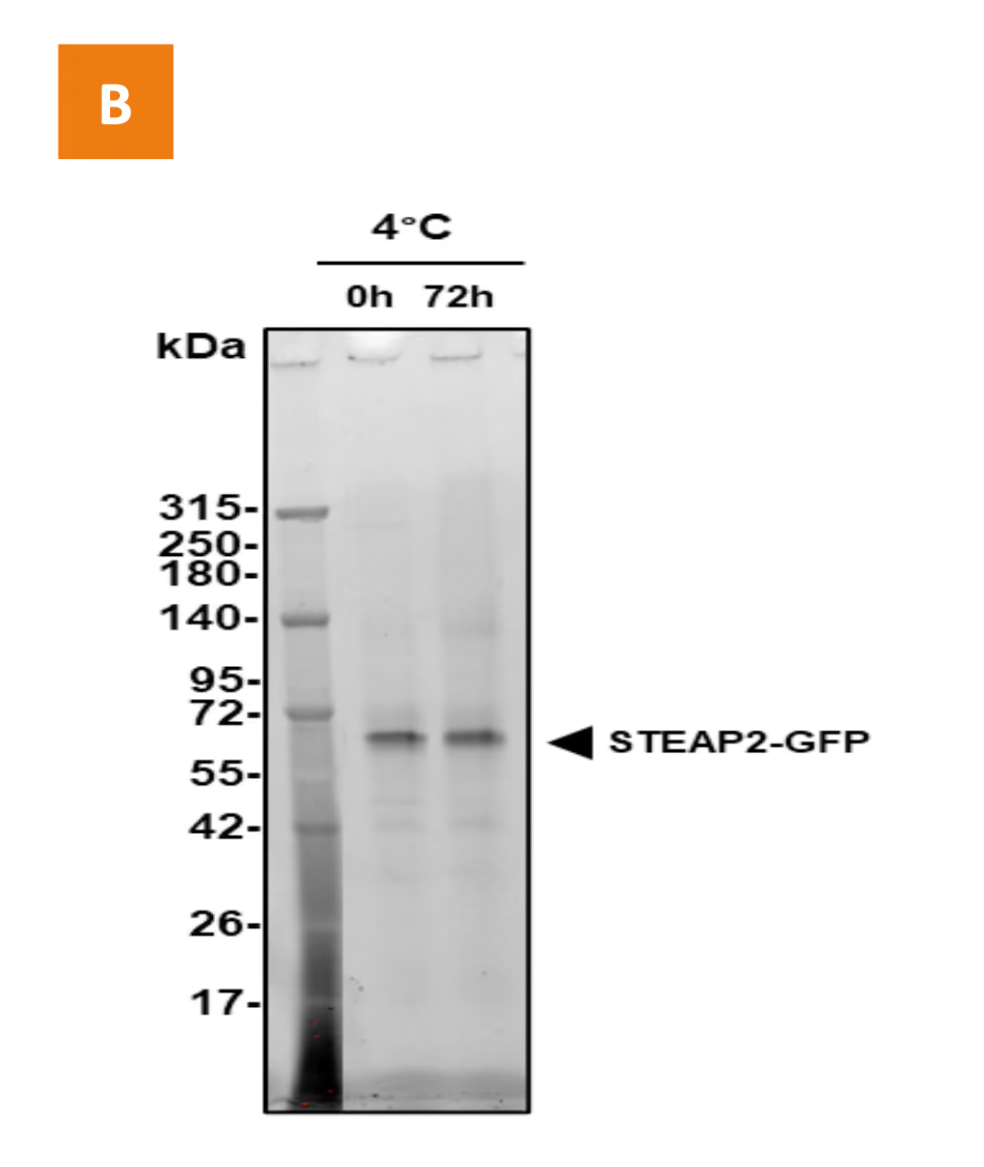
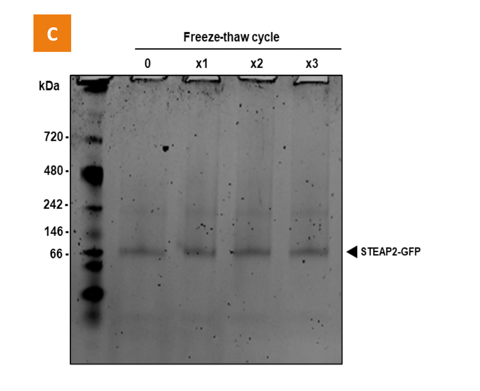
Figure 4. STEAP2 Purity and Stability. (A) SDS-PAGE analysis using Stain-Free™ detection. Full-length STEAP2 is indicated by the black arrow (approx. 80% purity). (B) Chrono-stability at 4°C. (C) Cryo-stability after freeze-thaw cycles.
Purified, Stable & Functional STEAP2
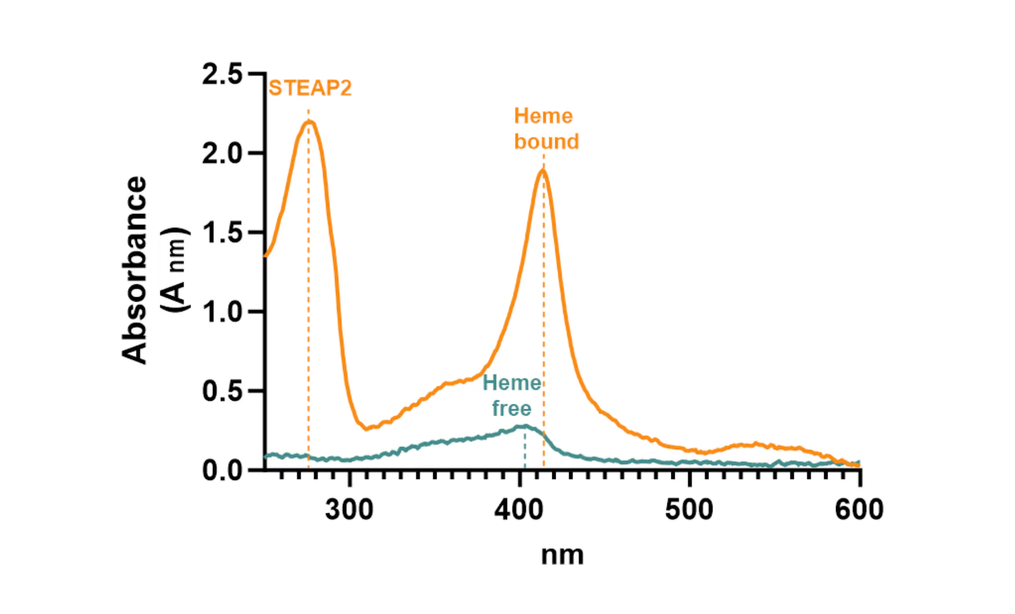
STEAP2 functionality was further assessed via heme-binding spectroscopy. As expected, a shift in absorbance from 403 nm (free heme) to 412 nm (bound heme) confirmed the heme-binding capacity and supported a native protein fold (Figure 5).
These findings confirm that the protein is both correctly folded and functionally active, providing a solid foundation for subsequent functional assays and structural investigations.
CryoEM Structure Determination
Cryo-electron microscopy (cryoEM) was used to confirm the identity, structural integrity, and ligand-binding mode of purified STEAP2. The workflow was divided into three successive stages: sample viability evaluation, high-resolution feasibility, and final structure determination.
Sample Viability Evaluation
Initial assessments were conducted to evaluate STEAP2 behavior under cryoEM at different concentrations and on multiple grid types and supports (Figure 6). Key quality indicators such as number of particles, orientational distribution, stability,stoichiometry, purity, and aggregation were monitored. A small dataset was acquired to evaluate particle distribution in micrographs, generate 2D class averages, and compute a low-resolution 3D map. This early-stage check allows discrepancies between biochemical QC and cryoEM visibility to be detected before committing to high-resolution efforts.
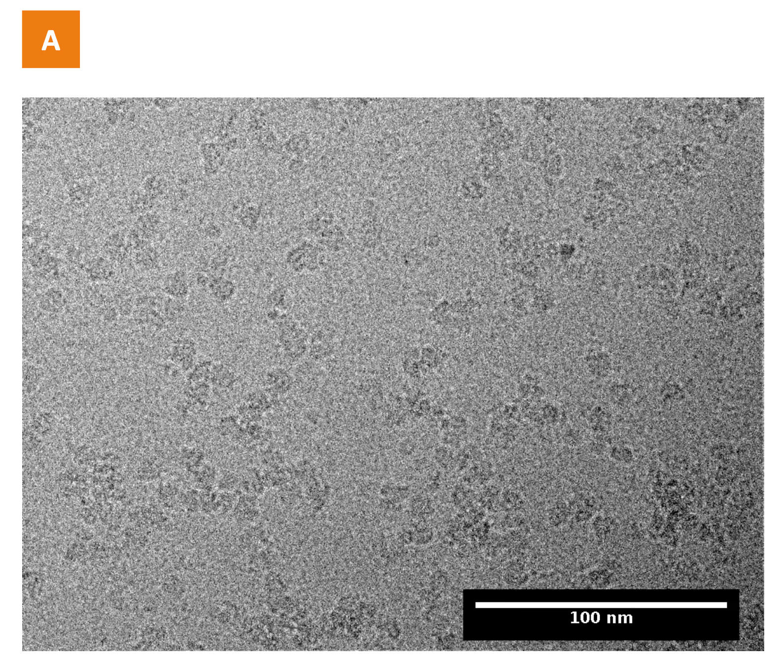
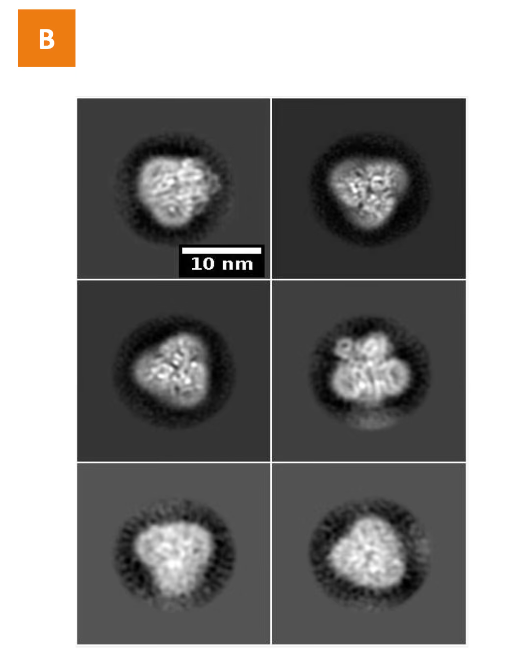
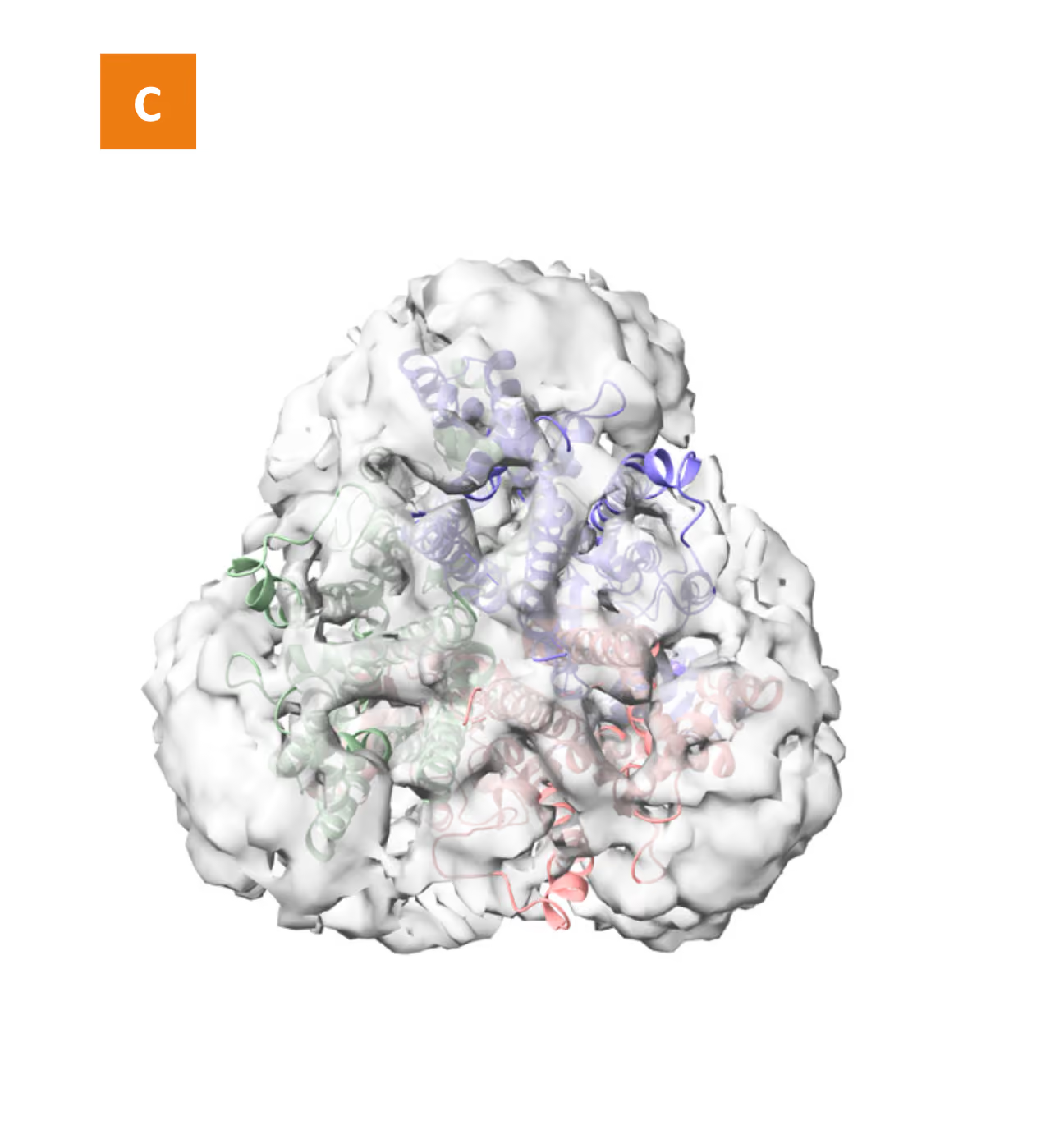
Figure 6. Small-scale cryoEM dataset of STEAP2. (A) Representative micrograph showing STEAP2 particles in vitreous ice. (B) 2D class averages revealing secondary structure and multiple orientations. (C) Low-resolution 3D map fitted with published structure (PDB: 7TAI), confirming sample identity.
High-Resolution Feasibility
Next, a dataset was collected to achieve a 3.5–4 Å resolution map, enabling rigid-body fitting and assessment of ligand presence (Figure 7). This step determines the presence or absence of preferred orientation, and if the protein can be reconstructed at higher resolution while determining the number of particles required to reach sub-3 Å resolution. Although small molecule ligands typically remain undetected at low resolutions due to their size and minimal conformational effects, this intermediate phase provides critical information before committing to large-scale data collection. Overall, this analysis provides the necessary confidence to move forward with in-depth structural exploration.
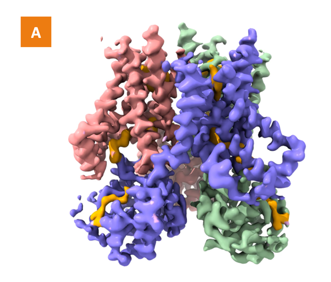
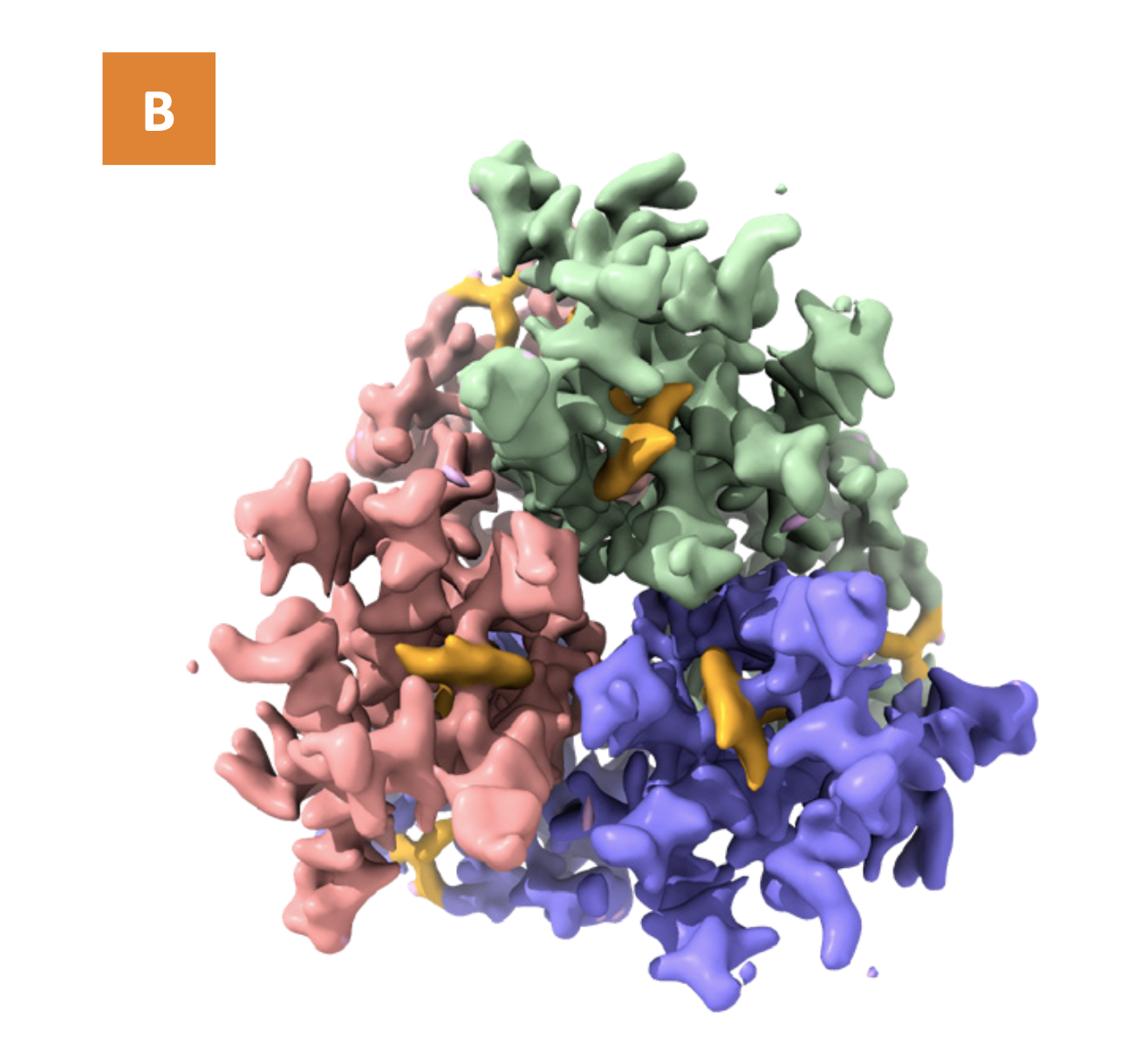
Figure 7. Middle-resolution map (~3.5 Å) of STEAP2 showing ligand density (orange) and favorable resolution scaling, supporting progression to final reconstruction. (A) Side view. (B) Top view, rotated 90° relative to (A).
High-Resolution Reconstruction and Model Building
In the final step, a full dataset was collected to achieve a 2.1 Å resolution map of the STEAP2 complex (Figure 8). The resulting map surpassed the resolution of the published structure (3.2 Å, PDB: 7TAI), allowing for high-precision model building.
The cryoEM map exhibited uniform resolution across the protein, enabling confident modeling of ligand-binding sites in transmembrane, extracellular, and intracellular regions.
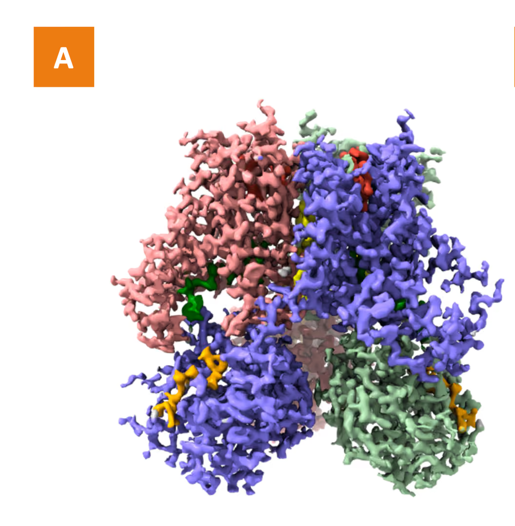
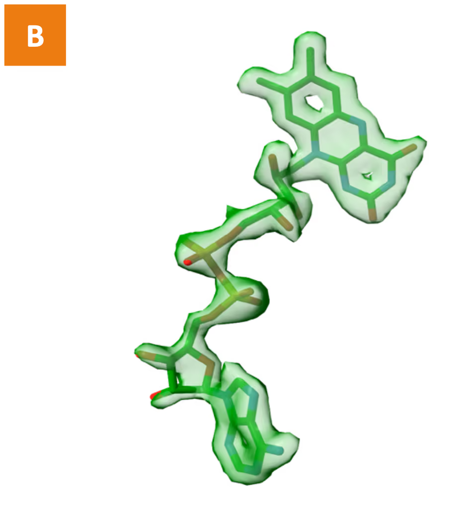
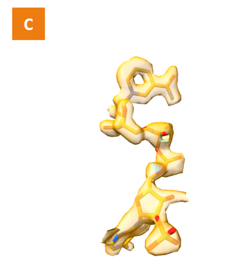
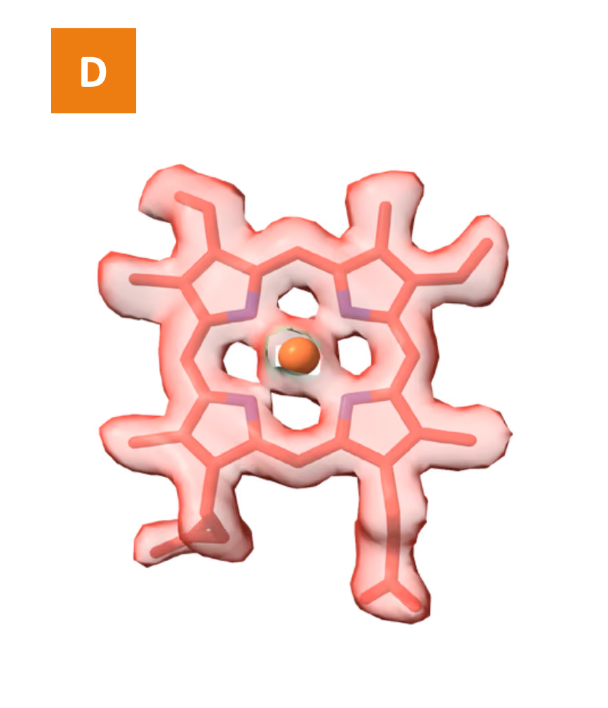
Figure 8. (A) Final 3D cryoEM map of STEAP2 at 2.1 Å resolution, color-coded by protein chains, ligands, and lipids. (B–D) High-confidence ligand models built at atomic resolution.
Additionally
protein–ligand interactions were analyzed in detail, with specific binding contacts visualized and annotated (Figure 9).
The reconstructed map enables detailed modelling of the entire pocket and all relevant interactions.
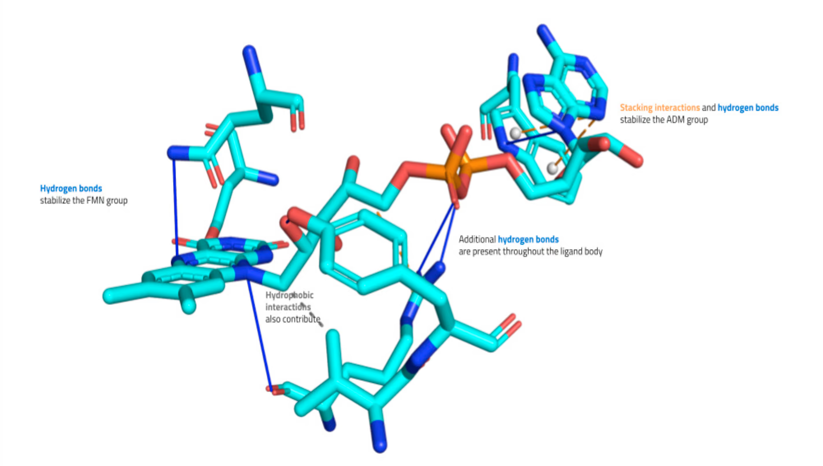
Figure 9. Representative STEAP2 ligand–protein interaction analysis highlighting key molecular contacts in the binding mode.
DISCUSSIONS
This study demonstrates the strength of the Eurofins CALIXAR platform in delivering high-quality recombinant membrane proteins suitable for advanced biophysical and structural studies. The STEAP2 protein was successfully produced in its native conformation, as confirmed through a series of rigorous biochemical assays, including ligand-binding, thermostability, and homogeneity analyses. These results underscore the ability of Eurofins CALIXAR to generate functional membrane proteins that retain native-like structure and ligand-binding properties highlighting the platform’s compatibility with downstream applications such as high-resolution cryo-EM. By combining Eurofins CALIXAR’s high-quality membrane protein production expertise with Eyen’s state-of-the-art cryo-electron microscopy (cryo-EM) capabilities, the platform provides an integrated, end- to-end solution—from gene design to high-resolution structural determination. This seamless “gene-to-structure” workflow enables efficient progression from early-stage protein production and validation to downstream cryo-EM analysis. On the structural front, the cryo-EM workflow developed in partnership with Eyen facilitates rapid evaluation of sample quality and ligand engagement, followed by the reconstruction of high-resolution 3D structures. In the case of STEAP2, this approach delivered a final map at 2.1 Å resolution—substantially improving upon the previously published 3.2 Å structure (PDB: 7TAI). This enhanced resolution is particularly valuable in small-molecule drug discovery, where atomic-level insights into ligand-binding interactions are often unattainable at resolutions above 3 Å.
Eurofins CALIXAR offers a scalable, reliable, and science-driven pathway for clients aiming to de-risk and accelerate structure- based drug design projects involving challenging membrane protein targets. Leverage Eurofins CALIXAR’s proprietary technology for producing highly pure, stable, and native-like recombinant proteins to enable high-resolution 3D co-structure determination of your lead–target complexes, and drive lead optimization through advanced computational chemistry.
References
- Torrez, C. Z. et al. STEAP2 promotes hepatocellular carcinoma progression via increased copper levels and stress-activated MAP kinase activity. Scientific Reports 14, (2024).
- Wang, X. et al. The copper-associated protein STEAP2 correlated with glioma prognosis and immune infiltration.Frontiers in Cellular Neuroscience 16, (2022).
- Wang, Y., Chen, S., Chen, S. & Jiang, J. Unveiling the role of copper metabolism and STEAP2 in idiopathic pulmonary fibrosis molecular landscape. Journal of Cellular and Molecular Medicine 28, (2024).
- Porkka, K. P., Helenius, M. A. & Visakorpi, T. Cloning and Characterization of a Novel Six-Transmembrane Protein STEAP2, Expressed in Normal and Malignant Prostate. Laboratory Investigation 82, 1573–1582.
- Gomes, I. M., Maia, C. J. & Santos, C. R. STEAP proteins: From structure to Applications in cancer therapy. Molecular Cancer Research 10, 573–587 (2012).
- Whiteland, H. et al. A role for STEAP2 in prostate cancer progression. Clinical & Experimental Metastasis 31, 909–920 (2014).
- Burnell, S. E. A. et al. STEAP2 knockdown reduces the invasive potential of prostate cancer cells. Scientific Reports 8, (2018).
- Jones, N. L. A. et al. Investigating STEAP2 as a potential therapeutic target for the treatment of aggressive prostate cancer. Cellular and Molecular Biology 69, 179–187 (2023).
- Yang, Q., Ji, G. & Li, J. STEAP2 is down-regulated in breast cancer tissue and suppresses PI3K/AKT signaling and breast cancer cell invasion in vitro and in vivo. Cancer Biology & Therapy 21, 278–291 (2019).
- Zanvit, P. et al. Antitumor activity of AZD0754, a dnTGFβRII-armored, STEAP2-targeted CAR-T cell therapy, in prostate cancer. Journal of Clinical Investigation 133, (2023).
- Chen, K. et al. Mechanism of stepwise electron transfer in six-transmembrane epithelial antigen of the prostate (STEAP) 1 and 2. eLife (2023) doi:10.7554/elife.88299.2.
